
Cell imaging is a vital technique for studying the structure, function, and dynamics of cells in various biological contexts. However, cell imaging can be challenging due to the complexity, diversity, and sensitivity of cellular samples, as well as the high demands for image quality, resolution, speed, and accuracy. Therefore, choosing the right microscope for cell imaging is crucial for obtaining reliable and meaningful results.
One of the most popular and widely used microscopes for cell imaging is the Evos microscope. The Evos microscope is a family of fully automated, multichannel fluorescence or brightfield microscopes that are designed by biologists for biologists. The Evos microscope offers a range of features and benefits that make it a versatile and powerful tool for cell imaging across a broad spectrum of applications.
In this article, we will introduce the Evos microscope, its features and benefits, its applications and sample data, its models and accessories, its usage and operation, its comparison with similar alternatives, and its conclusion.
What is Evos Microscope?
The Evos microscope is a product line of cell imaging systems developed by Thermo Fisher Scientific, a global leader in scientific products and services. The Evos microscope was launched in 2010 as a revolutionary alternative to conventional microscopes which are often bulky, complex, expensive, and difficult to use.
The Evos microscope is based on a modular design that allows users to customize their system according to their specific needs and preferences. The evos microscope consists of three main components:
- A high-resolution camera that captures stunning images in four-color fluorescence, transmitted light, or color modes.
- A bright and digitally controlled LED light source that provides uniform illumination across the entire field of view.
- Intuitive software that controls all aspects of image acquisition, processing, analysis, storage, and sharing.
The evos microscope also incorporates a compact and efficient design that saves space, energy, and costs. The evos microscope can be easily installed, maintained, aligned, calibrated, or upgraded without any external service or assistance. The Evos microscope can also be used within a biosafety cabinet or hood to ensure safety and sterility.
The evos microscope is exceptionally versatile and ideal for a wide range of cell imaging applications, such as fluorescence imaging, brightfield imaging, color imaging, live-cell imaging, cell counting and confluence measurements, Z-stacking and image stitching, time-lapse movies, multiwell plate scanning, wound healing assays, immunofluorescence staining, cell viability assays, apoptosis assays, transfection assays, gene expression assays, protein expression assays, drug screening assays, and many more.
The Evos microscope delivers superb images and data in no time, over time, every time at an exceptional value. The Evos microscope is easy to learn and operate and can revolutionize your cell imaging workflow.
Features and Benefits of Evos Microscope
The Evos microscope offers a number of features and benefits that make it a superior choice for cell imaging. Here are some of the main advantages of the Evos microscope:
High-resolution cameras and optics
The Evos microscope is equipped with high-resolution CMOS monochrome or color cameras that can capture images with up to 20 megapixels. The cameras have high sensitivity, low noise, fast frame rates, and large dynamic ranges, ensuring optimal image quality and resolution. The cameras also have a USB 3.0 interface that allows fast data transfer and storage.
The Evos microscope also has high-quality optics that provide clear and sharp images with minimal distortion or aberration. The Evos microscope has a range of objective lenses with different magnifications, numerical apertures, working distances, and correction types, allowing users to select the best lens for their application. The objective lenses are also color-coded and easy to change.
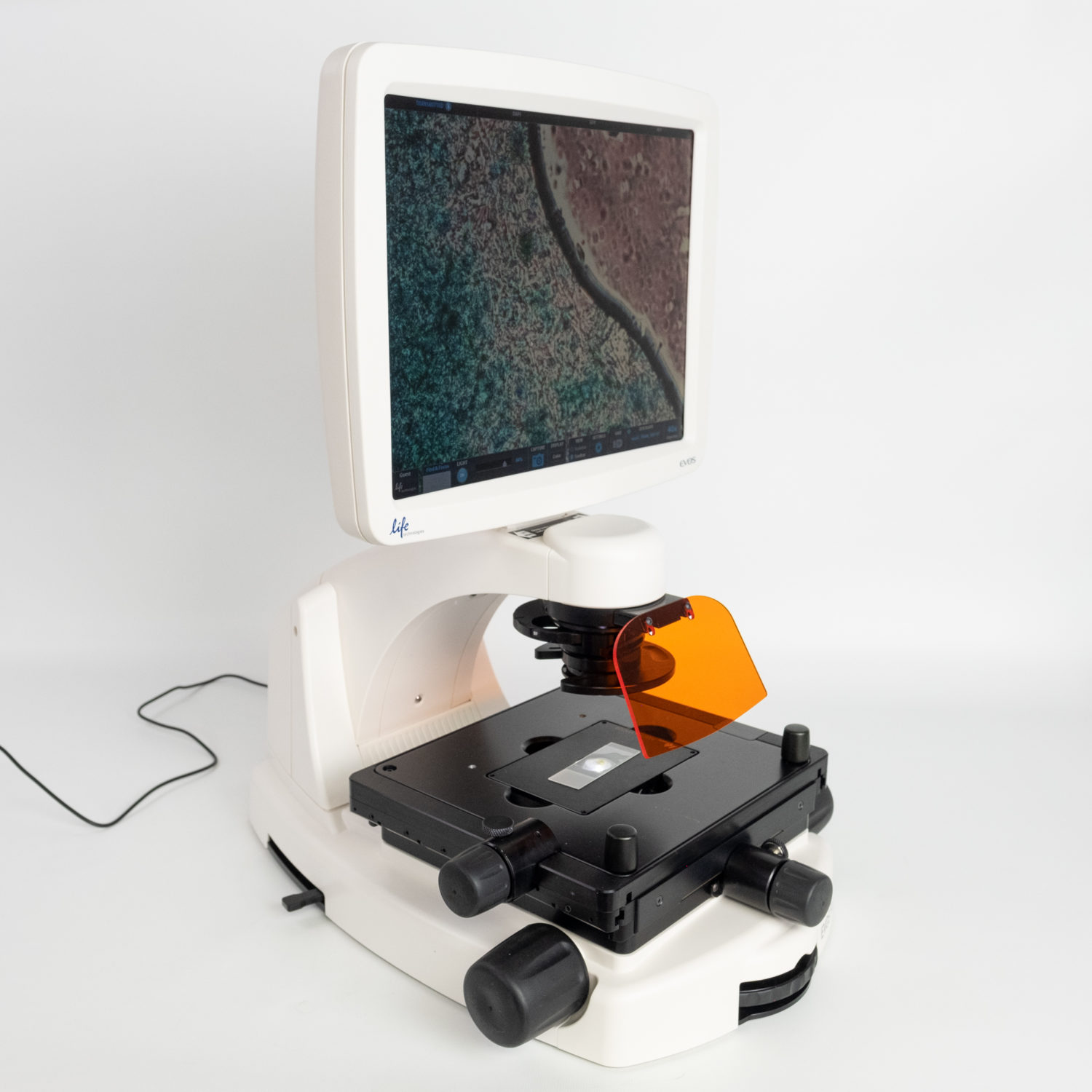
Bright and digitally controlled LED light sources
The Evos microscope uses LED light sources that are bright, stable, uniform, and long-lasting. The LED light sources have several advantages over traditional halogen or mercury lamps, such as:
- They consume less power and generate less heat, saving energy and costs.
- They have a longer lifespan and do not require replacement or maintenance, reducing downtime and waste.
- They have a lower risk of photobleaching or phototoxicity, preserving sample integrity and viability.
- They have a wider spectrum and can be tuned to specific wavelengths, enhancing fluorescence contrast and specificity.
- They can be switched on and off instantly, enabling fast image acquisition and analysis.
The Evos microscope has a modular design that allows users to choose from different LED light cubes that cover the most common fluorescence channels, such as DAPI, FITC, TRITC, Cy5, GFP, RFP, etc. The LED light cubes are easy to insert and remove and can be controlled digitally through the software. The software also allows users to adjust the intensity and duration of the LED light sources to optimize image quality and exposure.
Intuitive and user-friendly software
The Evos microscope has powerful and intuitive software that controls all aspects of image acquisition, processing, analysis, storage, and sharing. The software has a user interface designed by biologists for biologists, making it easy to learn and operate. The software has several features and tools that help users to capture stunning images in just a few clicks, such as:
- Autofocus: The software can automatically find the best focus for the sample using different methods, such as coarse or fine autofocus, manual focus, or focus lock.
- Z-stack: The software can automatically acquire images from different focal planes and assemble them into a single, optimally focused image.
- Time-lapse: The software can automatically capture images at regular intervals over a period of time and create a movie of the sample dynamics.
- Multiwell plate scanning: The software can automatically scan multiple wells of a multiwell plate and acquire images from each well.
- Image stitching: The software can automatically stitch together multiple images from adjacent fields of view to create a large image of the whole sample.
- Cell counting: The software can automatically count the number of cells in an image using different methods, such as thresholding or segmentation.
- Confluence measurements: The software can automatically measure the percentage of area covered by cells in an image using different methods, such as thresholding or segmentation.
- Image annotation: The software can add text, arrows, scale bars, or other annotations to the images to highlight important features or information.
- Image overlay: The software can overlay images from different channels or modes to create composite images that show multiple aspects of the sample.
- Image adjustment: The software can adjust the brightness, contrast, color, gamma, or other parameters of the images to enhance their appearance or visibility.
- Image export: The software can export the images in different formats, such as JPEG, PNG, TIFF, or BMP, or save them as PDF reports or PowerPoint presentations.
The Evos microscope software also has connectivity features that allow users to upload their images directly from the instrument to a Thermo Fisher Connect account. Thermo Fisher Connect is a cloud-based platform that enables users to access, manage, share, and analyze their data from anywhere, anytime, and on any device. Thermo Fisher Connect also offers additional post-acquisition image analysis tools through the EVOS Image Analysis application.
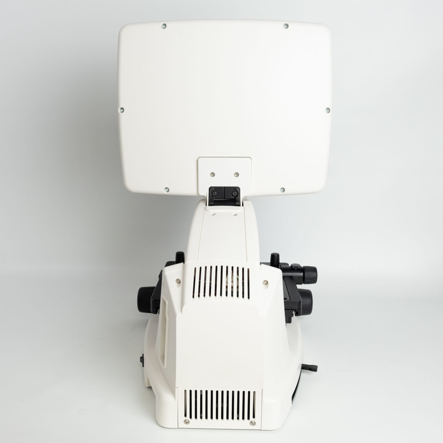
Compact and efficient design
The Evos microscope has a compact and efficient design that saves space, energy, and costs. The Evos microscope has a small footprint that fits on any benchtop or inside any biosafety cabinet or hood. The Evos microscope does not require any external components, such as power supplies, cooling fans, or filters, that may take up space or generate noise or heat. The Evos microscope also has a sleek and ergonomic design
that enhances user comfort and convenience. The Evos microscope has a large and high-definition LCD touchscreen that displays the images and the software interface. The Evos microscope also has a USB port that allows users to connect a mouse, keyboard, or flash drive for easier operation or data transfer.
Enhanced safety and minimal handling
The Evos microscope enhances user safety and minimizes sample handling. The evos microscope can be used within a biosafety cabinet or hood to ensure the safety and sterility of the samples and the users. The Evos microscope also has a sealed and easy-to-clean enclosure that prevents contamination or damage from spills or dust. The Evos microscope also has a UV light option that can be used to decontaminate the instrument after use.
The evos microscope also reduces the need for manual handling of the samples or the instrument. Most of the operational functions of the Evos microscope can be controlled through the software, eliminating the need to touch the instrument except to mount the slide and adjust the stage. The software also allows users to remotely control the Evos microscope from a computer or a mobile device via Thermo Fisher Connect. This reduces the risk of human error, sample loss, or sample degradation.
Applications and Sample Data of Evos Microscope
The Evos microscope is exceptionally versatile and ideal for a wide range of cell imaging applications. The evos microscope can capture images in four-color fluorescence, transmitted light, or color modes, allowing users to visualize different aspects of their samples, such as morphology, structure, function, expression, localization, interaction, or dynamics. The Evos microscope can also perform advanced imaging techniques, such as live-cell imaging, cell counting and confluence measurements, Z-stacking and image stitching, time-lapse movies, multiwell plate scanning, wound healing assays, etc.
Here are some examples of applications and sample data that can be obtained with the Evos microscope:
Fluorescence imaging
Fluorescence imaging is a technique that uses fluorescent molecules or probes to label specific molecules or structures in the sample and visualize them under a fluorescence microscope. Fluorescence imaging can reveal information about the identity, quantity, location, distribution, movement, or interaction of the molecules or structures of interest.
The Evos microscope can perform fluorescence imaging with high sensitivity, specificity, contrast, and resolution. The Evos microscope can capture images in four-color fluorescence mode using different LED light cubes that cover the most common fluorescence channels. The Evos microscope can also overlay images from different channels to create composite images that show multiple aspects of the sample.
For example, the image below shows a fluorescence image of HeLa cells stained with DAPI (blue) for nuclei, phalloidin (green) for actin filaments, and anti-tubulin antibody (red) for microtubules. The image was acquired with the EVOS M5000 Imaging System using DAPI/FITC/TRITC LED light cubes and a 40x objective lens.
Brightfield imaging
Brightfield imaging is a technique that uses transmitted light to illuminate the sample and visualize its morphology or structure under a brightfield microscope. Brightfield imaging can reveal information about the shape, size, texture, or density of the cells or tissues in the sample.
The evos microscope can perform brightfield imaging with high clarity, brightness, and contrast. The evos microscope can capture images in transmitted light mode using a white LED light source that provides uniform illumination across the entire field of view. The evos microscope can also adjust the brightness and contrast of the images to enhance their appearance or visibility.
For example, the image below shows a brightfield image of a mouse kidney tissue section stained with hematoxylin and eosin (H&E). The image was acquired with the EVOS M5000 Imaging System using a white LED light source and 20x objective lens.
Color imaging
Color imaging is a technique that uses color filters or cameras to capture images in color mode under a brightfield or fluorescence microscope. Color imaging can reveal information about the natural color or pigmentation of the cells or tissues in the sample, or enhance the contrast or visibility of certain features or structures in the sample.
The Evos microscope can perform color imaging with high fidelity, resolution, and speed. The Evos microscope can capture images in color mode using a high-resolution CMOS color camera that can produce images with up to 20 megapixels. The Evos microscope can also synthesize color images from monochrome images using different color filters or algorithms.
For example, the image below shows a color image of a mouse brain tissue section stained with cresyl violet. The image was acquired with the EVOS M5000 Imaging System using a color camera and 10x objective lens.
Live-cell imaging
Live-cell imaging is a technique that uses live-cell cultures to observe the behavior or dynamics of living cells under a microscope. Live-cell imaging can reveal information about the growth, division, movement, differentiation, communication, or response of the cells to various stimuli or conditions.
The Evos microscope can perform live-cell imaging with high stability, accuracy, and flexibility. The evos microscope can capture images of live-cell cultures in fluorescence or brightfield mode using different LED light sources that have a low risk of photobleaching or phototoxicity. The evos microscope can also perform time-lapse imaging to capture images at regular intervals over a period of time and create a movie of the cell dynamics.
The evos microscope can also be compatible with the EVOS Onstage Incubator, an optional accessory that provides precise control of temperature, humidity, and gases for live-cell analysis. The EVOS Onstage Incubator can maintain optimal environmental conditions for the live-cell cultures and allow users to monitor and adjust them remotely through the software.
For example, the image below shows a time-lapse movie of human umbilical vein endothelial cells (HUVECs) undergoing wound healing over 24 hours. The cells were stained with CellTracker Green CMFDA dye (green) for cytoplasm and Hoechst 33342 dye (blue) for nuclei. The movie was acquired with the EVOS M5000 Imaging System using FITC/DAPI LED light cubes and a 10x objective lens, and the EVOS Onstage Incubator set at 37°C, 5% CO2, and 95% humidity.
Cell counting and confluence measurements
Cell counting and confluence measurements are techniques that use image analysis to quantify the number or density of cells in an image. Cell counting and confluence measurements can reveal information about the viability, proliferation, or differentiation of the cells in the sample.
The evos microscope can perform cell counting and confluence measurements with high speed, accuracy, and reproducibility. The Evos microscope has onboard software that can automatically count the number of cells or measure the percentage of area covered by cells in an image using different methods, such as thresholding or segmentation. The software can also display the results as histograms, graphs, or tables, and export them as CSV files.
For example, the image below shows a cell counting result of HeLa cells stained with trypan blue dye. The software automatically counted the number of viable (unstained) and non-viable (stained) cells in the image using the thresholding method. The software also calculated the viability percentage as 88%. The image was acquired with the EVOS M5000 Imaging System using a white LED light source and a 10x objective lens.
Z-stacking and image stitching
Z-stacking and image stitching are techniques that use image processing to combine multiple images from different focal planes or fields of view into a single image. Z-stacking and image stitching can reveal information about the depth or width of the sample, or enhance the focus or resolution of the image.
The Evos microscope can perform Z-stacking and image stitching with high speed, quality, and automation. The Evos microscope has onboard software that can automatically acquire images from different focal planes or fields of view and assemble them into a single, optimally focused, or large image. The software can also adjust the alignment, blending, or cropping of the images to create a seamless and smooth image.
For example, the image below shows an image stitching result of a mouse intestine tissue section stained with H&E. The software automatically stitched together 16 images from adjacent fields of view to create a large image of the whole tissue section. The image was acquired with the
EVOS M5000 Imaging System using a white LED light source and 4x objective lens.
Models and Accessories of Evos Microscope
The Evos microscope has a modular design that allows users to choose from different models and accessories that suit their specific needs and preferences. The Evos microscope has six models and two accessories that offer different levels of performance, functionality, and compatibility. Here are the main features and differences of each model and accessory:
EVOS M5000 Imaging System
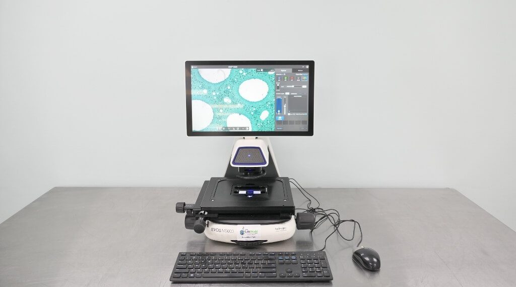
The EVOS M5000 Imaging System is the most advanced and versatile model of the evos microscope. The EVOS M5000 Imaging System offers the following features:
- A high-resolution CMOS monochrome camera that can capture images with up to 20 megapixels in four-color fluorescence, transmitted light, or color modes.
- A bright and digitally controlled LED light source that can accommodate up to four LED light cubes covering the most common fluorescence channels.
- A powerful and intuitive software that can perform advanced imaging techniques, such as autofocus, Z-stack, time-lapse, multiwell plate scanning, image stitching, cell counting, confluence measurements, image annotation, image overlay, image adjustment, image export, etc.
- A compact and efficient design that can fit on any benchtop or inside any biosafety cabinet or hood.
- An enhanced safety and minimal handling feature that allows most of the operational functions to be controlled through the software, eliminating the need to touch the instrument except to mount the slide and adjust the stage.
- A Thermo Fisher Connect connectivity feature that allows users to upload their images directly from the instrument to a Thermo Fisher Connect account, or download any new software upgrade directly in connected instruments.
- An EVOS Onstage Incubator compatibility feature that allows users to perform live-cell analysis with precise control of temperature, humidity, and gases.
- A Celleste Image Analysis Software compatibility feature that allows users to perform advanced image processing and analysis, such as 2D and 3D deconvolution and 3D visualization and analysis.
The EVOS M5000 Imaging System is ideal for users who need a high-performance, versatile, and user-friendly microscope for a wide range of cell imaging applications.
EVOS M7000 Imaging System
The EVOS M7000 Imaging System is a similar model to the EVOS M5000 Imaging System but with some differences. The EVOS M7000 Imaging System offers the following features:
- A high-resolution CMOS color camera that can capture images with up to 20 megapixels in four-color fluorescence or color modes.
- A bright and digitally controlled LED light source that can accommodate up to three LED light cubes covering the most common fluorescence channels.
- A powerful and intuitive software that can perform advanced imaging techniques, such as autofocus, Z-stack, time-lapse, multiwell plate scanning, image stitching, cell counting, confluence measurements, image annotation, image overlay, image adjustment, image export, etc.
- A compact and efficient design that can fit on any benchtop or inside any biosafety cabinet or hood.
- An enhanced safety and minimal handling feature that allows most of the operational functions to be controlled through the software, eliminating the need to touch the instrument except to mount the slide and adjust the stage.
- A Thermo Fisher Connect connectivity feature that allows users to upload their images directly from the instrument to a Thermo Fisher Connect account, or download any new software upgrade directly in connected instruments.
- An EVOS Onstage Incubator compatibility feature that allows users to perform live-cell analysis with precise control of temperature, humidity, and gases.
- A Celleste Image Analysis Software compatibility feature that allows users to perform advanced image processing and analysis, such as 2D and 3D deconvolution and 3D visualization and analysis.
The EVOS M7000 Imaging System is ideal for users who need a high-performance, versatile, and user-friendly microscope for a wide range of cell imaging applications, especially color imaging.
EVOS FL Auto 2 Imaging System
The EVOS FL Auto 2 Imaging System is an automated model of the evos microscope. The EVOS FL Auto 2 Imaging System offers the following features:
- A high-resolution CMOS monochrome camera that can capture images with up to 5 megapixels in four-color fluorescence or transmitted light modes.
- A bright and digitally controlled LED light source that can accommodate up to four LED light cubes covering the most common fluorescence channels.
- A powerful and intuitive software that can perform advanced imaging techniques, such as autofocus, Z-stack, time-lapse, multiwell plate scanning, image stitching, cell counting, confluence measurements, image annotation, image overlay, image adjustment, image export, etc.
- A compact and efficient design that can fit on any benchtop or inside any biosafety cabinet or hood.
- An enhanced safety and minimal handling feature that allows most of the operational functions to be controlled through the software, eliminating the need to touch the instrument except to mount the slide and adjust the stage.
- A Thermo Fisher Connect connectivity feature that allows users to upload their images directly from the instrument to a Thermo Fisher Connect account, or download any new software upgrade directly in connected instruments.
- An EVOS Onstage Incubator compatibility feature that allows users to perform live-cell analysis with precise control of temperature, humidity, and gases.
- A Celleste Image Analysis Software compatibility feature that allows users to perform advanced image processing and analysis, such as 2D and 3D deconvolution and 3D visualization and analysis.
- An automated stage and focus feature that allows users to scan large or multiple samples with high speed and accuracy.
- An automated filter wheel feature that allows users to switch between different fluorescence channels with high speed and precision.
The EVOS FL Auto 2 Imaging System is ideal for users who need a high-performance, automated, and user-friendly microscope for a wide range of cell imaging applications.
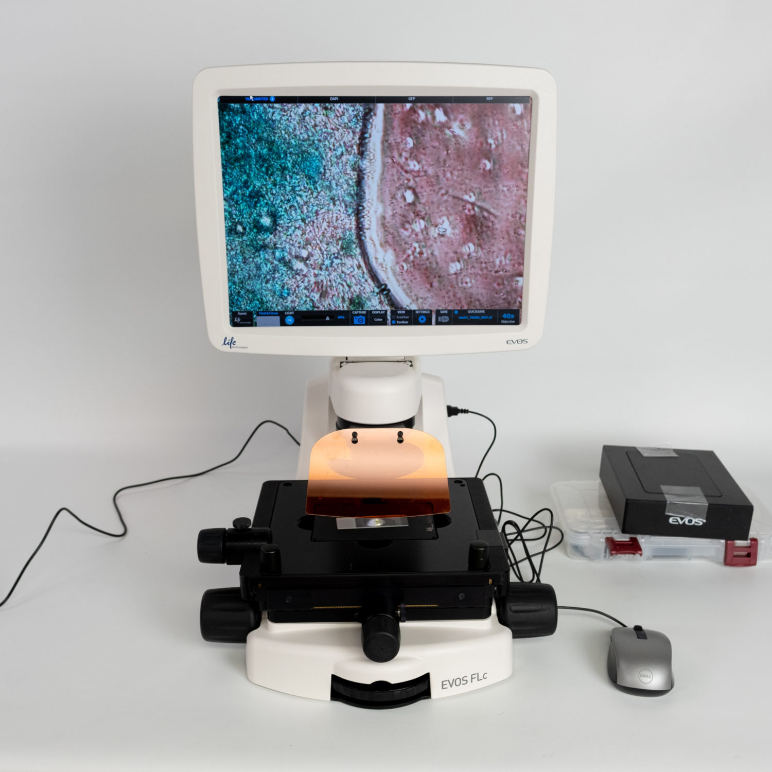
EVOS FLoid Cell Imaging Station
The EVOS FLoid Cell Imaging Station is a simple and affordable model of the evos microscope. The EVOS FLoid Cell Imaging Station offers the following features:
- A high-resolution CMOS color camera that can capture images with up to 5 megapixels in three-color fluorescence or color modes.
- A bright and digitally controlled LED light source that can accommodate up to three LED light cubes covering the most common fluorescence channels.
- Simple and easy-to-use software that can perform basic imaging techniques, such as autofocus, image capture, image overlay, image adjustment, image export, etc.
- A compact and efficient design that can fit on any benchtop or inside any biosafety cabinet or hood.
- An enhanced safety and minimal handling feature that allows most of the operational functions to be controlled through the software, eliminating the need to touch the instrument except to mount the slide and adjust the stage.
The EVOS FLoid Cell Imaging Station is ideal for users who need a simple, affordable, and user-friendly microscope for basic cell imaging applications.
EVOS XL Core Cell Imaging System
The EVOS XL Core Cell Imaging System is a large and robust model of the evos microscope. The EVOS XL Core Cell Imaging System offers the following features:
- A high-resolution CMOS color camera that can capture images with up to 5 megapixels in color mode.
- A bright and digitally controlled LED light source that provides uniform illumination across the entire field of view.
- Simple and easy-to-use software that can perform basic imaging techniques, such as autofocus, image capture, image adjustment, image export, etc.
- A large and robust design that can accommodate large or heavy samples, such as tissue sections, slides, dishes, flasks, or plates.
- An enhanced safety and minimal handling feature that allows most of the operational functions to be controlled through the software, eliminating the need to touch the instrument except to mount the slide and adjust the stage.
The EVOS XL Core Cell Imaging System is ideal for users who need a large, robust, and user-friendly microscope for basic color imaging applications.
EVOS Onstage Incubator
The EVOS Onstage Incubator is an optional accessory that can be compatible with the EVOS M5000, M7000, or FL Auto 2 Imaging Systems. The EVOS Onstage Incubator offers the following features:
- Precise control of temperature, humidity, and gases for live-cell analysis.
- Remote monitoring and adjustment of environmental conditions through the software.
- Compatibility with different types of samples, such as slides, dishes, flasks, or plates.
The EVOS Onstage Incubator is ideal for users who need to perform live-cell analysis with optimal environmental conditions.
Celleste Image Analysis Software
The Celleste Image Analysis Software is an optional accessory that can be compatible with any model of the Evos microscope. The Celleste Image Analysis Software offers the following features:
- An advanced image processing and analysis tool that can perform 2D and 3D deconvolution, 3D visualization and analysis, object segmentation and classification, colocalization analysis, intensity measurements, etc.
- A user-friendly interface that guides users through different steps of image analysis workflow.
- A customizable platform that allows users to create their own protocols or scripts for image analysis.
The Celleste Image Analysis Software is ideal for users who need to perform advanced image processing and analysis with their evos microscope images.
How to Use Evos Microscope?
The Evos microscope is easy to use and operate. Here are some general steps on how to use an Evos microscope:
Installation and Maintenance of Evos Microscope
The Evos microscope does not require any external service or assistance for installation or maintenance. The Evos microscope can be easily installed by following the instructions in the user manual or the online SmartStart guide. The Evos microscope can be easily maintained by performing some routine tasks, such as cleaning the enclosure, objective lenses, and LED light cubes with a soft cloth or lens paper, or decontaminating the instrument with a UV light option.
Workflow and Operation of Evos Microscope
The Evos microscope has a simple and intuitive workflow and operation. The evos microscope can be operated by following these steps:
- Turn on the evos microscope by pressing the power button on the back of the instrument.
- Select the desired LED light cube and an objective lens for your application. The LED light cube and objective lens are color-coded and easy to insert and remove.
- Mount your sample on the stage and adjust the stage position using the stage knobs or the software.
- Adjust the focus using the focus knob or the software. You can use different methods of autofocus, such as coarse or fine autofocus, manual focus, or focus lock.
- Adjust the brightness and exposure using the software. You can use different methods of exposure, such as auto exposure, manual exposure, or exposure lock.
- Capture your image using the software. You can use different modes of image capture, such as single image, multi-channel image, Z-stack image, time-lapse image, multiwell plate image, or stitched image.
- Process and analyze your image using the software. You can use different tools of image processing and analysis, such as image annotation, image overlay, image adjustment, cell counting, confluence measurements, etc.
- Save and share your image using the software. You can save your image in different formats, such as JPEG, PNG, TIFF, or BMP, or export your image as PDF reports or PowerPoint presentations. You can also upload your image directly from the instrument to a Thermo Fisher Connect account.
Image Acquisition and Analysis with Evos Microscope
The Evos microscope has powerful and intuitive software that can perform advanced image acquisition and analysis techniques. The Evos microscope software has several features and tools that help users to capture stunning images in just a few clicks, such as:
- Autofocus: The software can automatically find the best focus for the sample using different methods, such as coarse or fine autofocus, manual focus, or focus lock.
- Z-stack: The software can automatically acquire images from different focal planes and assemble them into a single, optimally focused image.
- Time-lapse: The software can automatically capture images at regular intervals over a period of time and create a movie of the sample dynamics.
- Multiwell plate scanning: The software can automatically scan multiple wells of a multiwell plate and acquire images from each well.
- Image stitching: The software can automatically stitch together multiple images from adjacent fields of view to create a large image of the whole sample.
- Cell counting: The software can automatically count the number of cells in an image using different methods, such as thresholding or segmentation.
- Confluence measurements: The software can automatically measure the percentage of area covered by cells in an image using different methods, such as thresholding or segmentation.
- Image annotation: The software can add text, arrows, scale bars, or other annotations to the images to highlight important features or information.
- Image overlay: The software can overlay images from different channels or modes to create composite images that show multiple aspects of the sample.
- Image adjustment: The software can adjust the brightness, contrast, color, gamma, or other parameters of the images to enhance their appearance or visibility.
- Image export: The software can export the images in different formats, such as JPEG, PNG, TIFF, or BMP, or save them as PDF reports or PowerPoint presentations.
The Evos microscope software also has connectivity features that allow users to upload their images directly from the instrument to a Thermo Fisher Connect account. Thermo Fisher Connect is a cloud-based platform that enables users to access, manage, share, and analyze their data from anywhere, anytime, and on any device. Thermo Fisher Connect also offers additional post-acquisition image analysis tools through the EVOS Image Analysis application.
The EVOS Image Analysis application is an optional accessory that can be compatible with any model of the evos microscope. The EVOS Image Analysis application is an advanced image processing and analysis tool that can perform 2D and 3D deconvolution, 3D visualization and analysis, object segmentation, and classification, colocalization analysis, intensity measurements, etc. The EVOS Image Analysis application has a user-friendly interface that guides users through different steps of the image analysis workflow. The EVOS Image Analysis application also has a customizable platform that allows users to create their own protocols or scripts for image analysis.
Comparison of Evos Microscope with Similar Alternatives
The Evos microscope is one of the most popular and widely used microscopes for cell imaging. However, there are also other similar alternatives that offer different features and benefits for cell imaging. Here are some of the main advantages and limitations of the evos microscope compared to other microscopes:
Advantages of Evos Microscope over Other Microscopes
The evos microscope has several advantages over other microscopes, such as:
- It is designed by biologists for biologists, making it easy to learn and operate.
- It has a modular design that allows users to customize their system according to their specific needs and preferences.
- It has a high-resolution camera that can capture images with up to 20 megapixels in four-color fluorescence, transmitted light, or color modes.
- It has a bright and digitally controlled LED light source that provides uniform illumination across the entire field of view and can be tuned to specific wavelengths.
- It has powerful and intuitive software that can perform advanced imaging techniques, such as autofocus, Z-stack, time-lapse, multiwell plate scanning, image stitching, cell counting, confluence measurements, image annotation, image overlay, image adjustment, image export, etc.
- It has a compact and efficient design that saves space, energy, and costs.
- It has an enhanced safety and minimal handling feature that allows most of the operational functions to be controlled through the software, eliminating the need to touch the instrument except to mount the slide and adjust the stage.
- It has a Thermo Fisher Connect connectivity feature that allows users to upload their images directly from the instrument to a Thermo Fisher Connect account, or download any new software upgrade directly in connected instruments.
- It has an EVOS Onstage Incubator compatibility feature that allows users to perform live-cell analysis with precise control of temperature, humidity, and gases.
- It has a Celleste Image Analysis Software compatibility feature that allows users to perform advanced image processing and analysis, such as 2D and 3D deconvolution and 3D visualization and analysis.
Limitations of Evos Microscope Compared to Other Microscopes
The evos microscope also has some limitations compared to other microscopes, such as:
- It does not have a confocal or super-resolution option that can provide higher resolution or contrast than conventional fluorescence microscopy.
- It does not have a phase contrast or differential interference contrast (DIC) option that can provide better visualization of unstained or transparent samples than brightfield microscopy.
- It does not have a darkfield or polarized light option that can provide better visualization of reflective or birefringent samples than brightfield microscopy.
- It does not have a scanning electron microscope (SEM) or transmission electron microscope (TEM) option that can provide higher magnification or resolution than optical microscopy.
Evos microscope evolution
The Evos microscope has evolved from previous models or releases to provide improvements, address issues, or otherwise help users in making a purchase decision. Here are some of the main changes and enhancements of the evos microscope over time:
- The Evos microscope was launched in 2010 as a revolutionary alternative to conventional microscopes which are often bulky, complex, expensive, and difficult to use.
- The Evos microscope was based on a modular design that allows users to customize their system according to their specific needs and preferences. The Evos microscope consists of three main components: a high-resolution camera, a bright and digitally controlled LED light source, and intuitive software.
- The Evos microscope has six models and two accessories that offer different levels of performance, functionality, and compatibility. The Evos microscope has the following models: EVOS M5000 Imaging System, EVOS M7000 Imaging System, EVOS FL Auto 2 Imaging System, EVOS FLoid Cell Imaging Station, EVOS XL Core Cell Imaging System, and EVOS Onstage Incubator. The Evos microscope also has the following accessories: Celleste Image Analysis Software and Thermo Fisher Connect.
- The Evos microscope has improved its camera resolution from 5 megapixels to 20 megapixels, allowing users to capture stunning images in four-color fluorescence, transmitted light, or color modes.
- The Evos microscope has expanded its LED light source options from three to four, covering the most common fluorescence channels, such as DAPI, FITC, TRITC, Cy5, GFP, RFP, etc. The Evos microscope also has a UV light option that can be used to decontaminate the instrument after use.
- The Evos microscope has enhanced its software features and tools that can perform advanced imaging techniques, such as autofocus, Z-stack, time-lapse, multiwell plate scanning, image stitching, cell counting, confluence measurements, image annotation, image overlay, image adjustment, image export, etc. The Evos microscope also has connectivity features that allow users to upload their images directly from the instrument to a Thermo Fisher Connect account, or download any new software upgrade directly in connected instruments.
- The Evos microscope has added an optional accessory that can provide precise control of temperature, humidity, and gases for live-cell analysis. The EVOS Onstage Incubator can maintain optimal environmental conditions for the live-cell cultures and allow users to monitor and adjust them remotely through the software.
- The Evos microscope has added an optional accessory that can perform advanced image processing and analysis, such as 2D and 3D deconvolution and 3D visualization and analysis. The Celleste Image Analysis Software can automatically quantify and analyze cellular images using different methods, such as object segmentation and classification, colocalization analysis, intensity measurements, etc.
These are some of the main improvements and enhancements of the evos microscope over time. The evos microscope is constantly evolving to meet the changing needs and expectations of the users. The Evos microscope is designed to eliminate the complexities of high-end microscopy without compromising performance. The Evos microscope is exceptionally versatile and ideal for a broad range of imaging applications, delivering superb images and data in no time, over time, every time at an exceptional value.
Evos microscope key choices and their effects on users.
When choosing an evos microscope, there are some important decision-making factors that users should consider. These factors include:
-
The type and mode of imaging:
Users should select the Evos microscope model that can capture images in the type and mode of imaging that they need for their application. For example, if users need to perform fluorescence imaging, they should choose a model that can accommodate LED light cubes for different fluorescence channels. If users need to perform color imaging, they should choose a model that has a color camera. If users need to perform live-cell imaging, they should choose a model that is compatible with the EVOS Onstage Incubator.
-
The resolution and quality of imaging:
Users should select the Evos microscope model that can capture images with the resolution and quality that they need for their application. For example, if users need to capture high-resolution images, they should choose a model that has a high-resolution camera with up to 20 megapixels. If users need to capture high-quality images, they should choose a model that has high-quality optics, bright and uniform LED light sources, and powerful and intuitive software.
-
The speed and automation of imaging:
Users should select the Evos microscope model that can capture images with the speed and automation that they need for their application. For example, if users need to capture images quickly, they should choose a model that has a fast frame rate, a USB 3.0 interface, and an auto exposure feature. If users need to capture images automatically, they should choose a model that has an automated stage and focus feature, an automated filter wheel feature, and an autofocus feature.
-
The versatility and compatibility of imaging:
Users should select the Evos microscope model that can capture images with the versatility and compatibility that they need for their application. For example, if users need to capture images of different types or sizes of samples, they should choose a model that has a range of objective lenses with different magnifications, numerical apertures, working distances, and correction types. If users need to capture images of different types or formats of data, they should choose a model that has a Thermo Fisher Connect connectivity feature or a Celleste Image Analysis Software compatibility feature.
These are some of the key choices and their effects on users when choosing an evos microscope. Users should weigh the pros and cons of each choice and select the Evos microscope model that best suits their needs and preferences. The Evos microscope is designed to offer a range of options and features that can cater to different user requirements and expectations. The Evos microscope is designed to provide users with stunning, easy-to-capture images that can help them achieve their research goals.
Evos Microscope Resources and Sellers
If you are interested in learning more about the Evos microscope or purchasing one, here are some useful resources and multiple sellers that you can check out:
- The official website of Thermo Fisher Scientific, the manufacturer of the Evos microscope, is where you can find detailed information about the features, models, accessories, sample data, services, and resources of the Evos microscope. You can also request a quote, a demo, or a contact from Thermo Fisher Scientific.
- The online store of Thermo Fisher Scientific, where you can browse and buy different models and accessories of the evos microscope. You can also compare prices, specifications, and reviews of different products.
- The online marketplace of LabX, where you can find new and used evos microscopes for sale from various sellers. You can also post a request for a quote or a product.
- The online marketplace of Lab Merchant, where you can find new and used evos microscopes for sale from various sellers. You can also post a request for a quote or a product.
These are some of the useful resources and multiple sellers on evos microscope that you can explore. You can also search for other sources or sellers on the internet or contact your local sales representative for more information. The Evos microscope is a versatile and powerful tool for cell imaging that can help you achieve your research goals.
Evos Microscope Reviews
When recommending Evos microscope options, it is helpful to provide first-hand evidence from users who have tried and tested the products. Here are some examples of user reviews that can support the recommendation of Evos microscope options:
- A user review of the EVOS FL and IRIS digital fluorescence microscopes by Sean Yu, a researcher at VitaScientific, a biotechnology company. The user compared the two systems in terms of instrument layout and design, image quality, feature comparison, and user experience. The user concluded that the EVOS FL is an excellent inverted microscope for routine cell culture usage, but is not designed to obtain the highest quality images. The user also praised the EVOS FL for its humanized design, easy operation, and powerful LED technology.
- A user review of the EVOS Cell Imaging Systems by Peter Drain, a professor at the University of Pittsburgh School of Medicine. The user highlighted the advantages of the EVOS over traditional microscopes, such as its humanized design, widefield fluorescent images, integrated intuitive design, and powerful LED technology. The user also shared how his lab benefits from the EVOS all-in-one design.
- A user review of the EVOS FL Auto Imaging System by Ruchika Nadav Nongram, a researcher at Queen Mary University of London. The user shared her experience of using the EVOS FL Auto for cardiac repair and heart failure research. The user appreciated the EVOS FL Auto for its ease of use, automation, image quality, and versatility. The user also mentioned how the EVOS FL Auto helped her to save time and resources.
- A user review of the EVOS FL Auto Imaging System by Lucia Cordero Espinosa, a researcher at the University of Cambridge. The user shared her experience of using the EVOS FL Auto for liver regeneration research. The user praised the EVOS FL Auto for its high-resolution images, fast scanning, and live-cell imaging capabilities. The user also mentioned how the EVOS FL Auto helped her to obtain reliable and reproducible data.
These are some examples of first-hand evidence that can support the recommendation of evos microscope options. Users can also search for more reviews or testimonials on the internet or contact their local sales representative for more information. The Evos microscope is a versatile and powerful tool for cell imaging that can help users achieve their research goals.
Conclusion
The Evos microscope is a versatile and powerful tool for cell imaging across a broad range of applications. The Evos microscope offers a range of features and benefits that make it a superior choice for cell imaging. The Evos microscope is easy to learn and operate and can revolutionize your cell imaging workflow.
If you are looking for a high-performance, versatile, and user-friendly microscope for cell imaging, the Evos microscope is the right choice for you.
Frequently Asked Questions
Here are some frequently asked questions about the Evos microscope:
How much does the Evos microscope cost?
The price of the Evos microscope depends on the model and accessories you choose. You can request a quote from Thermo Fisher Scientific or contact your local sales representative for more information.
How can I get technical support or service for the Evos microscope?
You can get technical support or service for the Evos microscope by contacting Thermo Fisher Scientific through phone, email, or online chat. You can also visit the Thermo Fisher Scientific website for more resources, such as user manuals, videos, webinars, protocols, etc.
How can I get training or education on how to use the Evos microscope?
You can get training or education on how to use the evos microscope by attending online or onsite courses offered by Thermo Fisher Scientific. You can also visit the Thermo Fisher Scientific website for more resources, such as user manuals, videos, webinars, protocols, etc.
How can I get more information or updates on the evos microscope?
You can get more information or updates on the Evos microscope by visiting the Thermo Fisher Scientific website or following Thermo Fisher Scientific on social media platforms, such as Facebook, Twitter, LinkedIn, YouTube, etc.

